Retinoschisis is a retinal condition in which the layers of the retina separate or split from each other. It can sometimes resemble a retinal detachment (RD) during an eye examination, but the two conditions are different.
In retinal detachment, the neurosensory retina separates from the underlying retinal pigment epithelium (RPE). In retinoschisis, the separation occurs within the retinal layers themselves.
At Soni Eye Care & Super Speciality Retina Clinic in Mulund, Mumbai, retinoschisis can be evaluated with a detailed retinal examination and appropriate imaging such as OCT, fundus photography, perimetry and B-scan ultrasonography when required. The clinic provides specialised medical and surgical retina care.
What is Retinoschisis?
Retinoschisis occurs when the retinal layers split apart, creating a space within the retina. It may occur as an inherited condition, particularly in children and young males, or may develop as a degenerative change in older adults.
Retinoschisis is broadly classified into:
- Degenerative or acquired retinoschisis – more commonly seen in adults and often discovered incidentally during a dilated retinal examination.
- Inherited retinoschisis – may occur as part of inherited retinal disorders such as X-linked juvenile retinoschisis (XLRS).
- Macular retinoschisis – when the splitting involves the central retina or macula, it can affect vision.
Some inherited retinal disorders, including Goldmann-Favre syndrome, can also be associated with retinoschisis-like changes.
Retinoschisis vs Retinal Detachment
Retinoschisis and retinal detachment can appear similar, but they are not the same condition.
In retinal detachment, the neurosensory retina separates from the RPE. Retinal detachment can threaten vision and may require urgent treatment.
In retinoschisis, the retina splits within its own layers. Many cases, particularly degenerative retinoschisis, remain stable and do not require treatment.
Because the distinction can sometimes be difficult, a detailed examination by a vitreoretina specialist is important when retinoschisis or retinal detachment is suspected.
What are the Symptoms of Retinoschisis?
Symptoms depend on the type and location of retinoschisis.
Degenerative Retinoschisis
Degenerative retinoschisis is frequently asymptomatic. It is often discovered incidentally during a dilated retinal examination.
Some patients may notice:
- A dark or missing area in part of the visual field
- Floaters
- Reduced peripheral vision
- Visual changes if the macula becomes involved
Flashes are not a typical feature of uncomplicated retinoschisis. However, new flashes, a sudden increase in floaters or a curtain/shadow in the vision should be evaluated urgently, because these symptoms can occur with retinal tears or retinal detachment.
Juvenile or Inherited Retinoschisis
X-linked juvenile retinoschisis usually presents in childhood or young adulthood and can cause:
- Reduced vision
- Distortion or metamorphopsia
- Difficulty seeing fine details
- Changes involving the macula
- Abnormalities on OCT examination
The severity can vary considerably between individuals.
How is Retinoschisis Diagnosed?
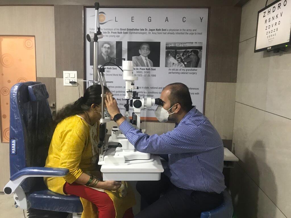
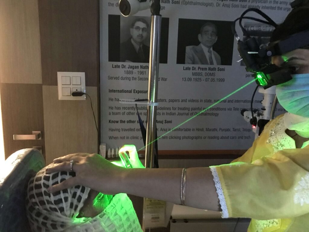
A comprehensive retinal examination is the first step in diagnosing retinoschisis.
Depending on the patient’s age, symptoms and examination findings, the retina specialist may recommend:
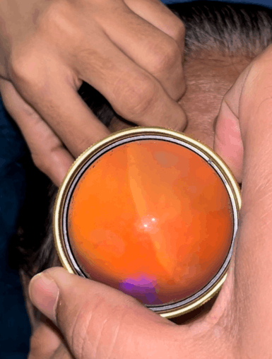
Dilated Retinal Examination
A detailed examination of the peripheral retina helps identify the characteristic appearance of retinoschisis and distinguish it from retinal detachment, retinal tears and other peripheral retinal conditions.
Optical Coherence Tomography (OCT)
OCT is particularly useful for macular retinoschisis and juvenile retinoschisis. It provides high-resolution cross-sectional images of the retinal layers and can demonstrate splitting within the retina.
Fundus Photography
Fundus photographs provide documentation of retinal findings and can be useful for monitoring changes over time.
B-Scan Ultrasonography
A B-scan may be recommended when the retina cannot be adequately visualised, such as when there is media opacity, vitreous haemorrhage or another condition obscuring the view. It can also help differentiate retinal pathology from other causes of an elevated retina.
Visual Field Testing / Perimetry
Perimetry may be useful in selected patients to evaluate peripheral visual-field defects or areas of reduced retinal function.
Genetic Evaluation
In children or young patients with suspected inherited retinoschisis, particularly when there is a family history, genetic counselling and genetic testing may be considered.
Does Retinoschisis Require Treatment?
Not every patient with retinoschisis needs treatment.
Treatment depends on the type of retinoschisis, whether the macula is involved, the patient’s vision and whether complications such as retinal detachment or significant vitreoretinal traction are present.
Observation
Most cases of uncomplicated degenerative retinoschisis can be observed without surgery when vision is stable and there are no sight-threatening complications.
Periodic retinal examinations may be recommended to monitor the condition.
Treatment of Juvenile Retinoschisis
Management of inherited or juvenile retinoschisis depends on the extent of retinal involvement and visual impairment.
In selected patients with macular involvement, topical carbonic anhydrase inhibitors such as dorzolamide or brinzolamide may be considered by the treating retina specialist. Their response can vary between patients.
If significant vitreoretinal traction or another surgical indication is present, vitreoretinal surgery may be considered.
Surgery for Complicated Retinoschisis
Surgery is generally reserved for selected cases with complications, such as a retinal detachment associated with retinoschisis or significant traction affecting vision.
Depending on the retinal findings, surgical options may include:
- Pars plana vitrectomy
- Scleral buckle surgery
- Combination retinal surgery in selected cases
The appropriate procedure depends on the location and extent of the retinal pathology and should be decided after a detailed retinal examination.
Is Retinoschisis Dangerous?
Most cases of degenerative retinoschisis are relatively stable and do not lead to serious visual loss.
However, a retinal examination is important because retinoschisis can sometimes coexist with retinal breaks or retinal detachment, and distinguishing between retinoschisis and retinal detachment is clinically important.
If you suddenly develop:
- Flashes of light
- A sudden increase in floaters
- A curtain or shadow over your vision
- Sudden loss of peripheral vision
- Sudden reduction in vision
you should seek an urgent retinal evaluation.
Retinoschisis Treatment in Mumbai
If you have been diagnosed with retinoschisis or have been told that you may have retinoschisis versus retinal detachment, a consultation with a vitreoretina specialist in Mumbai can help establish the diagnosis and determine whether observation or treatment is required.
Soni Eye Care & Super Speciality Retina Clinic, Mulund, Mumbai provides specialised retinal evaluation and treatment, with facilities including OCT, fundus photography, perimetry, B-scan ultrasonography, retinal lasers, intravitreal treatment and vitreoretinal surgery.
The clinic is located in Mulund West, Mumbai, and provides access to specialised retina care for patients from Mulund, Thane, Kalyan, Navi Mumbai and other parts of Mumbai.
Retina Specialists at Soni Eye Care
Dr. Anuj Soni, DO, DNB (LVPEI), FICO, Fellow Vitreo Retina, FAICO, has a special interest in retinal disorders including retinal detachment, diabetic retinopathy, age-related retinal disorders and inflammatory retinal conditions.
Dr. Asmita Soni is a vitreoretinal specialist practising at Soni Eye Care in Mulund West, with interests including vitreoretinal surgery, retinal lasers, intravitreal treatment and management of retinal disorders.
Why Choose Soni Eye Care for Retinoschisis Evaluation?
- Dedicated super-speciality retina clinic in Mulund
- Consultation with vitreoretina specialists
- OCT and retinal imaging facilities
- Fundus photography
- Perimetry / visual-field testing
- B-scan ultrasonography
- Retinal laser facilities
- Vitreoretinal surgical services
- Evaluation of retinal detachment and other retinal disorders
Soni Eye Care’s official clinic information lists its Mulund centre at Rohini Residency, M.G. Road, near Panch Rasta, Mulund West, Mumbai – 400080.
Frequently Asked Questions About Retinoschisis
Is retinoschisis the same as retinal detachment?
No. In retinoschisis, the retinal layers separate from each other. In retinal detachment, the neurosensory retina separates from the RPE. The two conditions can sometimes look similar, which is why specialist retinal examination is important.
Can retinoschisis cause blindness?
Most cases of degenerative retinoschisis do not cause severe vision loss. However, inherited retinoschisis involving the macula or retinoschisis complicated by retinal detachment can significantly affect vision.
Does degenerative retinoschisis need surgery?
Usually not. If vision is stable and there are no significant complications, observation is often appropriate. Surgery is considered when there are specific complications or a significant threat to vision.
Can retinoschisis be treated with eye drops?
In selected cases of juvenile or inherited retinoschisis with macular involvement, topical carbonic anhydrase inhibitors such as dorzolamide may be considered. Treatment should be individualised by a retina specialist.
Can retinoschisis turn into retinal detachment?
Retinoschisis itself is different from retinal detachment. However, retinal breaks or retinal detachment can occur in association with retinoschisis in some patients, which is why periodic retinal evaluation may be recommended.
Which doctor should I see for retinoschisis?
A vitreoretina specialist or retina surgeon is the appropriate specialist for evaluating suspected retinoschisis, particularly when there is a question of retinal detachment, retinal breaks, macular involvement or visual-field loss.
Book a Retina Consultation in Mulund, Mumbai
If you have been diagnosed with retinoschisis, have peripheral retinal changes, or have been advised to rule out retinal detachment, consult a vitreoretina specialist for a detailed retinal examination.
Soni Eye Care & Super Speciality Retina Clinic
Mulund West, Mumbai
Call to schedule an appointment: +91 77159 45144